Researcher Spotlight Series


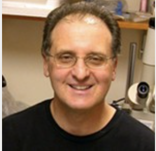
Douglas Marchuk, PhD Daniel Snellings
In this Researcher Spotlight, we spoke with Doug Marchuk, PhD, and Daniel Snellings from the Duke University School of Medicine. Doug and Daniel’s work centers on the genetics that underlies cardiovascular diseases. They recently published a paper in Nature called “PIK3CA and CCM Mutations Fuel Cavernomas Through a Cancer-like Mechanism.” Doug and Daniel chatted with us about their exciting findings regarding the genetics of cerebral cavernous malformations.
Brittany Enzmann: Can you tell us about your research on cardiovascular disease and how you became interested in this field?
Doug Marchuk: I have been interested in the vascular aspect of cardiovascular phenotypes and specifically what are often called vascular malformations or vascular anomalies. What intrigued me about these was the fact that you have these extremely abnormal vascular structures in the context of otherwise normal vasculature. I was very interested in picking apart the genes that caused these. That’s when I’ve been doing that for 28 years now.
Brittany: You published in Nature on cerebral cavernous malformations, also called CCMs. Can you explain more about what this disorder is and what questions you wanted to answer?
Doug: Cerebral cavernous malformation or some people call it a cavernoma, is a vascular malformation. The disease is named after the malformation itself. They are clusters of dilated vessels that occur in the brain, so they are cerebral. They’re cavernous, that is, they are hugely dilated vessels with defective junctional components. They’re malformed vessels, but they’re very discrete lesions.
They cause either a slow leak or oozing or hemorrhage or can overtly bleed. They cause cerebral events, like seizures or hemorrhagic stroke. They are fairly common as a lesion on autopsy studies. If you just take brains and you look, you may find one or two of these, but they generally would have been asymptomatic, But there are occasional sporadic cases where there’s a very large one. Dan will talk about that in a minute about why we think those big ones occur. Then in the familial forms, these patients often have dozens to even over a hundred of these in their brain because they have a predisposition. It is an autosomal dominant phenotype. We were involved along with a French lab identifying genes involved in the syndrome in terms of the inherited form. So, what I was interested in is understanding why, if a patient has a germline mutation and is predisposed to this, did he get these discrete individual lesions where the vasculature just a few centimeters away is completely normal? What was causing the actual formation as opposed to just the predisposition? That leads to some of the studies that Dan and others have done in my lab and other labs as well.
Brittany: You note that the disease mechanism of CCMs is analogous to cancer. What findings led you to this conclusion?
Doug: That Dan’s work, so take it away Dan.
Daniel Snellings: Thanks, Doug. So, previous studies had noted that a lot of these CCMs form as a result of a somatic mutation in one of three different genes implicated in familial CCM. And it is that somatic mutation event that initiates these lesions. One of the things that couldn’t be fully explained by this phenomenon was that some CCMs are far worse than others. As Doug said, a lot of these sporadic CCMs tend to be very large and very aggressive, whereas you also have very large and aggressive CCMs in familial disease. But you also have hundreds of these small, sometimes microscopic lesions that just are along for the ride. So, what is differentiating between these small lesions and these large lesions? We hypothesized that there could be mutations in other genes that act as a modifier where a mutation in gene A could result in a very large CCM and mutation in gene B could do something else and it could make CCMs more aggressive or less aggressive.
We carried out a sequencing study of about 80 CCMs and we found quite surprisingly that about 80% of the CCMs we sequenced had a somatic gain-of-function mutation in a gene called PI3 kinase, particularly the catalytic subunit of PI3 kinase. This was really surprising because PI3 kinase is a gene that’s commonly mutated in cancers. I believe after TP53, it’s the second most mutated gene across all cancers. CCM is a discretely not-cancer disease — compared to cancer being very aggressive, very metastatic. Aspects that we associate with cancer CCM doesn’t fulfill. So, it’s quite surprising to find a mutation in a cancer gene.
We had these previously discovered somatic mutations in a set of CCM genes and this new mutation in PI3 kinase. What’s interesting is we have several CCMs that have both these CCM gene mutations and PI3 kinase mutations. It turns out that this evokes a model similar to cancer, where we have a biallelic mutation of this vascular suppressor gene, just like tumor suppressor genes. Then we have gain-of-function of this vascular activating gene, just like oncogenes. So, although phenotypically CCMs are very different than cancers, genetically there is a lot of overlap.
Brittany: As part of your study, you investigated the genetic architecture of the CCMs using single-cell DNA sequencing. What did you find and why was it important to investigate this disease at the single-cell level?
Daniel: We had found that there are multiple different somatic mutations in these CCMs —some of them in these CCM genes and some of them in PI3 kinase.
The critical question at that point was whether or not all of these different somatic mutations were happening in the same cells or whether or not these were distinct populations of cells that kind of came together to form a mixed malformation. This has really big implications for cell autonomy and whether or not these mutations are synergizing, or they have completely distinct roles in the pathogenesis of CCM. So, we needed to figure out whether or not these were in the same cell. The way to do that is with single-cell DNA sequencing, and because these mutations are quite rare — oftentimes less than 5% of the alleles carry these mutations — using more traditional approaches like sorting individual cells into plates and amplifying the genome weren’t feasible.”
“We needed a targeted high-throughput method to sequence these cells. Tapestri was really the perfect way to do that. We were able to easily isolate nuclei from frozen CCMs and run them through the Tapestri Platform to amplify the CCM genes and PI3 kinase. We were able to figure out that in all of the cases the CCM mutations and the PI3 kinase mutations happened in the same cells.”
This continued into mechanistic studies where our collaborators at the University of Pennsylvania showed that PI3 kinase activation feeds into the CCM signaling cascade so that these mutations are having a synergistic effect, which validates why these mutations need to happen in the same cell.
Brittany: Now that you’ve identified that these aggressive CCMs have this three-hit mechanism, so in the CCM genes and then in the PI3 kinase gene and what would you like to investigate next?
Daniel: One of the really big questions is whether or not somatic large variants, like structural variants, contribute to CCM pathogenesis. We’ve been able to detect really small point mutations like SNPs and indels, but there are a lot of CCMs where we still haven’t found somatic mutations. Beyond CCM, we also have diseases like hereditary hemorrhagic telangiectasia and other vascular diseases. And beyond vascular diseases— like cancer— we have really great sensitivity for picking up low-frequency small variants. But the sensitivity is very poor for picking up large variants that could cause loss of heterozygosity and contribute to the pathogenesis of the disease.
Our immediate experimental ideas are to use the Tapestri Platform to identify regions of loss of heterozygosity that are present in a very small group of cells as few as 10 or 20 cells. Then, we would observe if there is overlap in known areas of pathogenic contribution — to see whether or not loss of heterozygosity is contributing to disease. Beyond that, I think that there are a lot of larger biological questions that this kind of science can address. Doug will talk a little bit about that.
Doug:
“One of the beauties of the Tapestri system is the DNA part. We’re doing single nucleus in our case, but also single-cell single DNA analysis is possible. There are other platforms for doing RNA, but we really needed to look at the DNA. I think what’s very clear that it’s not only in cancer but in a lot of diseases, there are somatic mutations. If they’re at low enough frequency, you may or may not pick them in bulk tissue. To understand the genotype of individual cells — what different variants are in there— you need to do it at the single-cell or single-nucleus level.”
I’m just really excited about pushing this all the way. As Dan mentioned, this whole concept of loss of heterozygosity, for example — a very large deletion — would not be picked up if it’s in low frequency. And yet, in an individual cell, it’s either there or not there. So, we’re really excited about pushing and limit and probing the genetic variation. Each individual cell may have a different genotype and you need to look at the single-cell DNA level to determine that. I’m really excited about that.
There are other aspects of what the Mark Kahn Lab at Penn and our lab have discovered that relate to therapeutics, which we can talk about another time. Of course, that’s the end game. It’s exciting to see mutations and things, but we really want to help the patients. I think we’re on our way with that too.
Brittany: Thank you so much, Doug and Daniel, for chatting with us today, we really look forward to your future work on CCMs and other vascular diseases.
Read Doug and Daniel’s Nature paper here!


